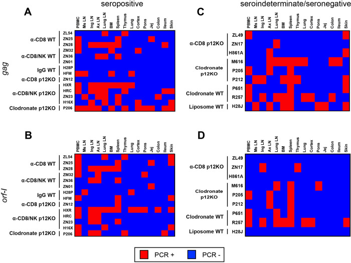
Fig 3. Viral dissemination in infected animals.
(A-D) Heat map of the nested PCR amplifying the gag and orf-I genes in biopsies from the ileum, colon, thymus, skin, jejunum, cortex, lung, spleen, inguinal LN, mesenteric LN, axillary LN, and lung LN, collected at the time of euthanasia following virus inoculation, together with PBMCs and bone marrow for (A,B) seropositive and (C,D) sero-indeterminate/seronegative animals. Genomic DNA was extracted to amplify viral DNA. Nested PCR was performed using primers designed to amplify the gag and orf-I genes (see Materials and Methods). The orf-I PCR products were sequenced to verify HTLV-1p12KO virus versus HTLV-1WT. Positive amplification of either (A,C) gag or (B,D) orf-I by nested PCR is shown in red; absence of amplification is shown in blue.

