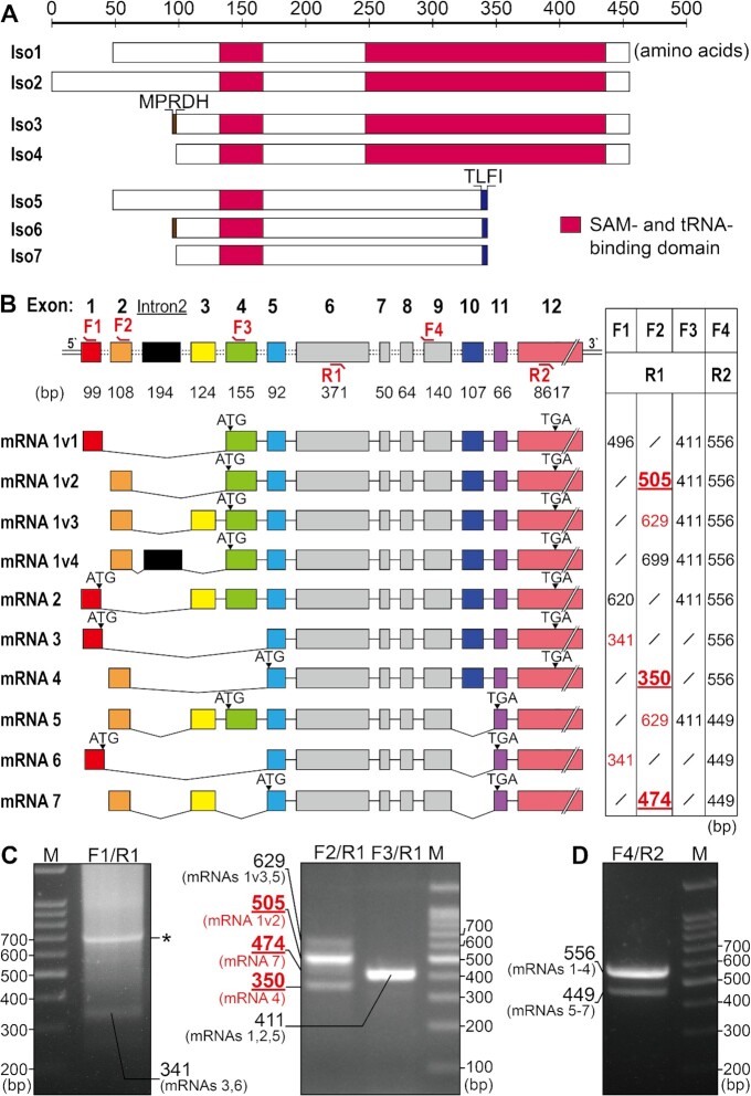
Figure 1.
Protein and mRNA analyses of METTL8. (A) Schematic showing NCBI-annotated METTL8 protein isoforms. Isoforms 5–7 lack a complete SAM and tRNA binding domain. Isoforms 3 and 4 differ only in the presence of five residues (MPRDH) in the former. The same difference exists between isoforms 5 and 6. A TLFI motif in isoforms 5–7, which was absent in isoforms 1–4 is also indicated. (B) Schematic showing NCBI-annotated METTL8 mRNA variants with each exon indicated. The black box represents intron 2. mRNAs 1v1-4 encoded the identical METTL8-Iso1 protein isoform, while mRNAs 2–7 encoded isoforms METTL8 Iso2-7. The pairing positions of primers used for mRNA identification are labeled at the top of the relevant exons. Bands that were successfully amplified using F1 and F2 forward primers and DNA sequenced are highlighted in red, while predicted bands not obtained by PCR are marked in black. (C, D) Agarose gel images of PCR products with different primer combinations as indicated. Each lane was confirmed by DNA sequencing. A nonspecific band not derived from the METTL8 gene is marked by an asterisk in (C). Three bold and underlined sizes represent DNA products that could be definitely assigned to specific mRNA isoforms in (B) and (C).

