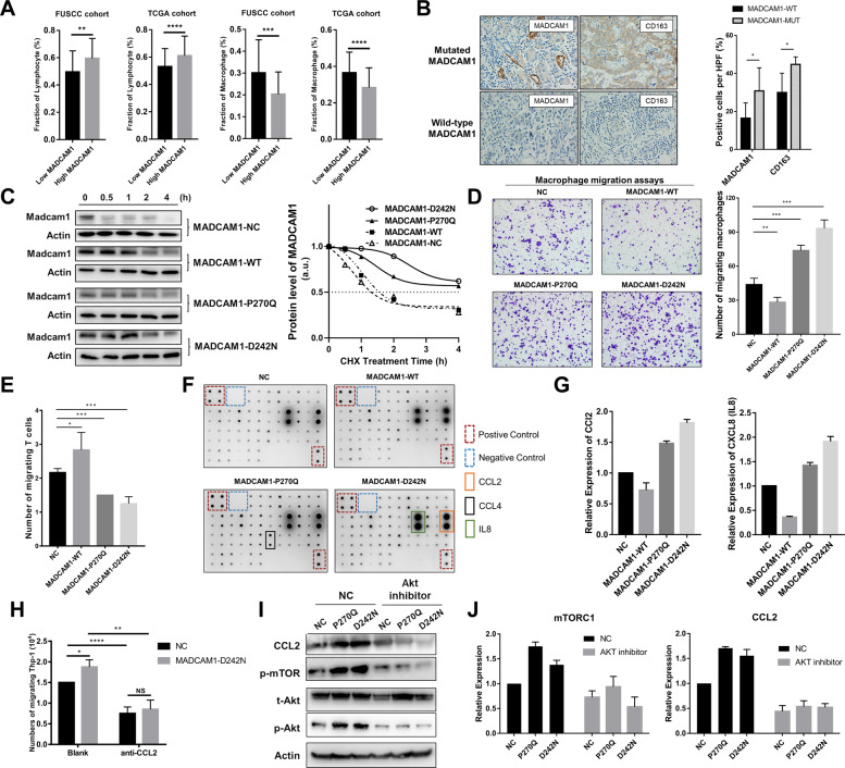
Fig. 5. MADCAM1 mutants associates with chemotactic migration of T cells and M2 macrophages.
A The expression of MADCAM1 show a significant positive correlation with the fraction of lymphocyte and a negative correlation with the fraction of macrophages, in both TCGA cohort and FUSCC cohort. B Immunohistochemistry (IHC) analysis showed that the pericellular staining intensity of MADCAM1 in the tumor cells of samples carrying mutated MADCAM1 was significantly stronger than those carrying wild-type MADCAM1. Additionally, CD163 was significantly positively stained in the infiltrating immune cells of samples with mutated MADCAM1, when compared with those with wild-type MADCAM1. C Effect of MADCAM1P270Q and MADCAMD242N on stability of the Madcam1 protein. GC cells transfected vectors expressing wild-type MADCAM1 (MADCAM1-WT), MADCAM1P270Q mutants (MADCAM1-P270Q), MADCAM1D242N mutants (MADCAM1-D242N) or negative control (MADCAM1-NC) were treated with 100 μg/ml cycloheximide (CHX) and then were harvested for western blot at indicated time points. Western blot data are quantified by ImageJ software. D Macrophage migration assay by the supernatants from MADCAM1WT, MADCAM1MUT and control GC cells. E T cell migration assay by the supernatants from MADCAM1WT, MADCAM1MUT and control GC cells. F The chemokine profiles of corresponding supernatants detected by Human Chemokine Antibody Arrays. G Expression of CCL2 and IL8 in supernatant from MADCAM1WT, MADCAM1MUT and control GC cells. H Blockading CCL2 by anti-CCL2 antibody reduced migration of macrophages, especially in MADCAM1D242N GC cells. I The p-Akt, CCL2 and p-mTOR was blocked by Akt inhibitor Perifosine, while the total levels of Akt were hardly changed. J The expression of p-mTOR and CCL2 was obviously suppressed after inhibition of Akt in MADCAM1P270Q and MADCAM1D242N GC cells. Bars, ±SD; *p < 0.05; **p < 0.01. ***p < 0.001.

