FIGURE 6.
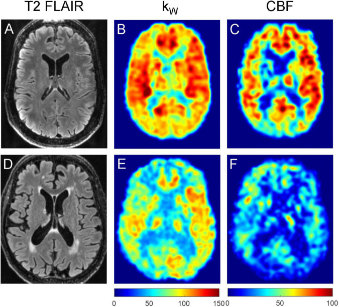
Representative T2-FLAIR images (A,D), kW maps (B,E) and CBF maps (C,F) obtained from a 76-year-old subject with no vascular risk factors and no significant WMH (A–C), and a 74-year-old subject with two vascular risk factors (HTN, HLD) and moderate burden of WMH (D–F). The subject without appreciable WMH demonstrated mean cortical kW of 98.4 min–1, mean white matter kW of 93.8 min–1, mean cortical CBF of 56.6 mL/100 g/min, and mean white matter CBF of 47.6 mL/100 g/min. The subject with moderate WMH burden (total WMH volume of 5,456 mm3) demonstrated lower mean cortical kW of 72.6 min–1, mean white matter kW of 64.0 min–1, mean cortical CBF of 28.1 mL/100 g/min, and mean white matter CBF of 23.6 mL/100 g/min.

