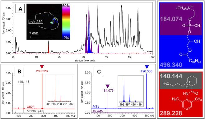
FIGURE 3.
LC-MS and LC-MS/MS analyses of non-volatile components of serum collected from a lung cancer patient. (A) XICs of intact molecular ions at m/z 496.3 (blue) and m/z 289.2 (red), and a fragment ion at m/z 184.1 (purple) plotted on the background of the total ion chromatogram (shown with a black trace). The MS and MS/MS datasets shown at the bottom of this panel were collected at elution time of 15 min (B) and 32 min (C), allowing these two species to be identified as bupivacaine and LPC16:0, respectively. The structures of both molecular ions and the most abundant fragments alongside their calculated masses are shown in the right-hand-side diagram. The inset in panel A shows a spatial distribution of the bupivacaine signal within the cross-section of a biopsy tissue obtained with MALDI MSI.

