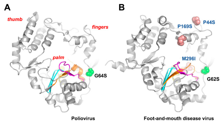
Figure 3.
Poliovirus (A) and foot-and-mouth disease virus (B) RdRp structures showing the location of motifs A–D and residues relevant for ribavirin binding and resistance. Locations of motifs A, B, C, and D are shown in orange, yellow, blue, and magenta, respectively [34,121]. Spheres are used to represent the location of relevant amino acid substitutions. Crystal structures were taken from PDB files 2ILY (poliovirus RdRp) and 1U09 (foot-and-mouth disease virus RdRp).

