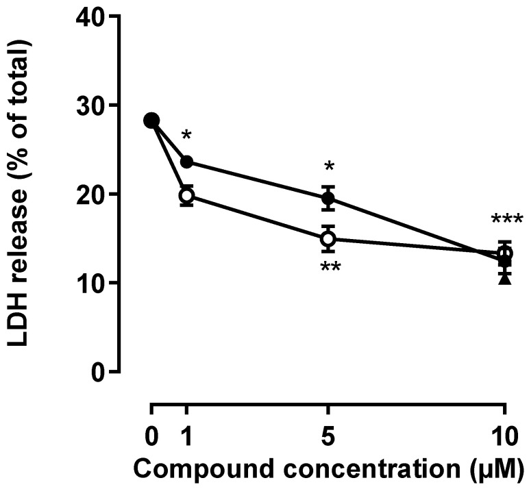
Figure 1.
Phosphorous dendrimers prevent NMDA-mediated excitotoxicity. Neurons were treated with the indicated concentration of phosphorous dendrimers G3 (open circles), G4 (closed circles), or MnTBAP (10 µM; closed triangle) for 1 h; and then NMDA (150 µM) was added and incubation continued for another 24 h. LDH activity was determined as indicated in the Materials and Methods section. Zero concentration represents net (stimulated–basal) NMDA-induced LDH release in the absence of any other treatment. The data represent the mean ± the s.e.m. of 6 to 12 independent experiments. * p < 0.05, ** p < 0.01, and *** p < 0.001 when compared to NMDA-treated cells.

