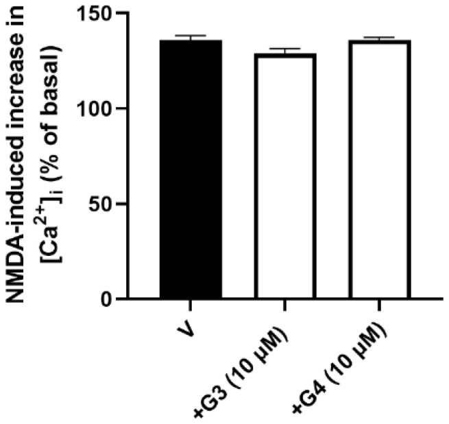
Figure 2.
Effect of phosphorous dendrimers on NMDA-induced increase in [Ca2+]i. Neurons were treated (10 µM; 1 h) with G3 or G4 phosphorous dendrimers. Then, the neurons were incubated with Fura-2 and exposed to NMDA (150 µM) in the absence (V) and presence of dendrimers. Data represent the percentage over basal line (100%) of the ratio of fluorescence taken as an index of [Ca2+]i, as indicated in the Materials and Methods section. The data are expressed as the mean + the s.e.m. of 30 to 40 neurons obtained from three independent experiments.

