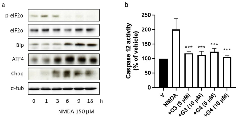
Figure 5.
Effect of phosphorous dendrimers on NMDA-induced changes in ER-stress-signaling proteins and in caspase 12 activity. (a) Neurons were treated with vehicle (V) or the indicated concentration of G3 or G4 phosphorous dendrimers for 1 h. Then, NMDA (150 µM) was added and the incubation continued for another 1 h for p-eiF2α and eiF2α determination and for 6 h for Bip, ATF4, and Chop protein determination by Western blot, as described in the Materials and Methods section. α-Tubulin was used as loading control. The image shows one representative experiment that was repeated three times with similar results. (b) Neurons were treated with vehicle (V) or the indicated concentration of phosphorous dendrimers for 1 h. Then, NMDA (150 µM) was added and after 6 h, caspase 12 activity measured as indicated in the Methods section. The data represent the mean + the s.e.m. of three experiments. *** p < 0.001 as compared to NMDA-treated cells in the absence of dendrimers.

