Figure 3.
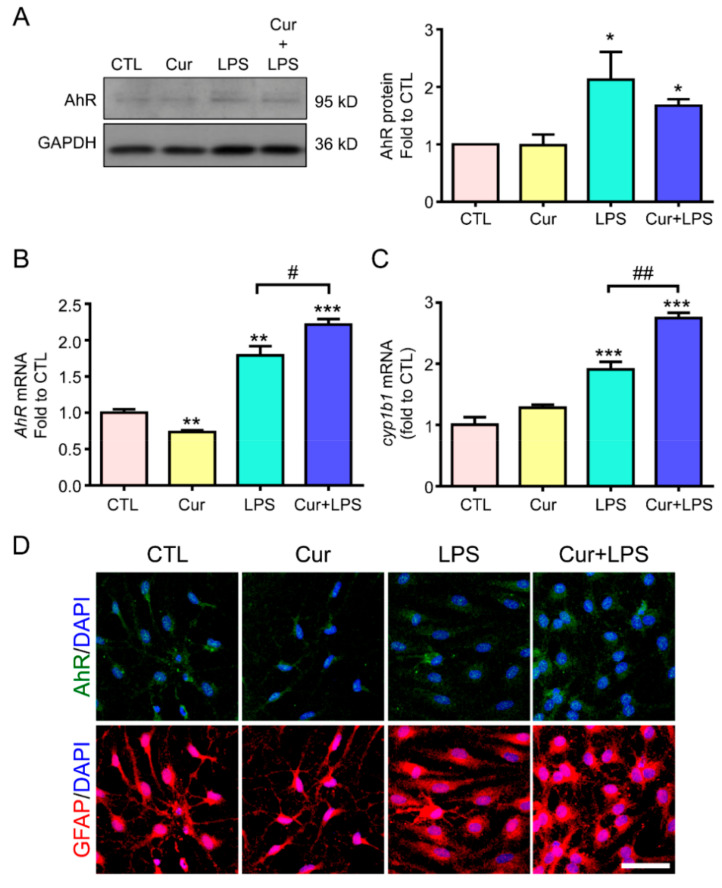
Curcumin enhanced LPS-induced AhR activation and nuclear translocation in astrocytes. Primary astrocytes were treated with 1 µg/mL LPS, 1 µM curcumin (Cur), or their combined treatment for 12 h in RNA and 24 h in protein. (A) AhR protein expression was determined by western blotting. GAPDH was used as the loading control. A representative blot and the quantification results are shown. n = 3. (B,C) AhR (B) and CYP1B1 (C) mRNA expression was determined by RT-PCR. Results are presented as fold expression differences over the control condition. (D) Immunofluorescence was performed to evaluate the expression of AhR (green, upper panel) and GFAP (an astrocyte marker; red, lower panel); DAPI (blue) was used to counterstain the nuclei. Representative images are shown. Note that curcumin enhanced LPS-induced AhR mRNA, not protein level, and elevated LPS-increased CYP1B1 mRNA expression and AhR nuclear translocation in astrocytes. Scale Bar = 20 μm. Statistical differences were analyzed using the unpaired t-test, and represented as: * p < 0.05, ** p < 0.005, *** p < 0.001 compared with the control group and # p < 0.05, ## p < 0.01 compared with the LPS group. DAPI, 4′,6-diamidino-2-phenylindole.

