Figure 3.
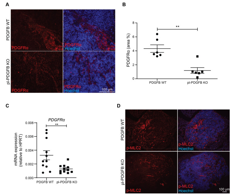
Fewer cancer-associated fibroblasts and reduced myosin light chain (MLC) phosphorylation in tumors from pl-PDGFB KO mice. (A,B) Tumor sections from 14-week old WT and pl-PDGFB KO RT2-positive mice were immunostained for PDGFRα (n = 6/group, ** p = 0.0022) and the positive area quantified using Image J. (C) Expression of PDGFRα at the transcriptional level was analyzed by qPCR using RNA extracted from the same type of tumors as in panel A and B (n = 11/group, ** p = 0.0095). (D,E) Tumor sections from 14-week old WT and pl-PDGFB KO RT2-positive mice were immunostained for phosphorylated MLC2 (p-MLC2) (WT n = 7; KO n = 8, * p = 0.0184), and the positive areas quantified using Image J. (F) Co-staining of PDGFRα and p-MLC was performed on tumor tissue from 14-week old RT2-positive mice WT and pl-PDGFB KO mice. Error bars in the graphs represent the standard error of mean (SEM).


