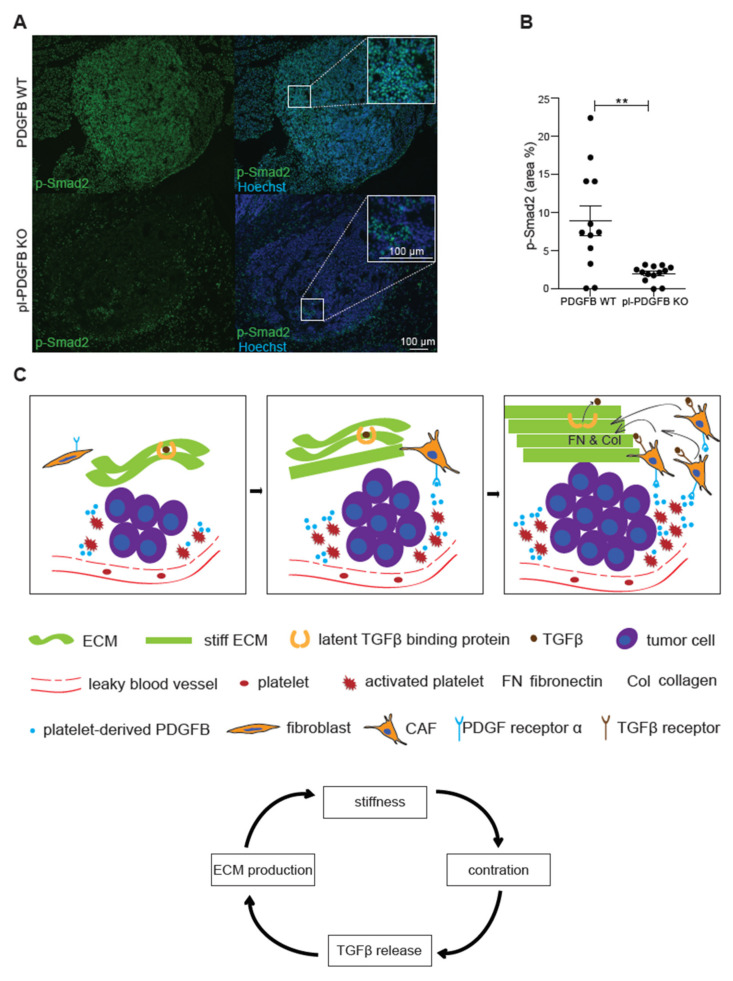
Figure 4.
Reduced TGFβ signaling in tumors from mice lacking PDGFB in platelets. (A,B) Tumors from 14-week old WT and pl-PDGFB KO RT2-positive mice were immunostained for p-Smad2 and the positive areas were quantified using Image J (WT n = 12; KO n = 13, ** p = 0.0015). Error bars in the graphs represent the standard error of mean (SEM). (C) Schematic model: the discontinuous endothelium in a tumor exposes platelets to subendothelial spaces, leading to activation, degranulation and release of PDGFB and other platelet-derived molecules in the tumor microenvironment (left panel). The PDGFRα+ CAFs are recruited by PDGFB derived from platelets and other sources in the tumor microenvironment. CAFs express large amounts of ECM proteins such as Col1 and FN, which leads to increased stiffness of the ECM (middle panel). Increased cell contraction due to the stiffer ECM leads to more release of active TGFβ from its extracellular stores, which in turn leads to more ECM expression and subsequently more FN polymerization (right panel). This process is creating a vicious circle (the arrow flow chart at the bottom): the more ECM production, the stiffer matrix and more release of active TGFβ, a potent inducer of ECM expression.

