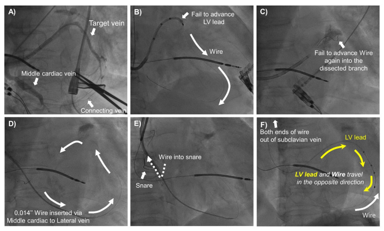
Figure 3.
The Antidromic Snare Technique. (A) Venography showed the target vein (lateral) and the connecting vein to the middle cardiac vein. (B) Failure to advance the LV lead into the target vein. (C) Re-wiring into the target branch was unsuccessful due to the dissected ostium of the target vein. (D) The 0.014″ guidewire was advanced into the middle cardiac vein and back into the CS ostium via the connecting vein (white arrow). (E) The guidewire distal end was snared at the right atrium. (F) The distal end of the guidewire was snared out of the sheath. The LV lead was advanced into the target vein via the guidewire distal end (yellow arrow). The LV lead and guidewire traveled in opposite directions. CS, coronary sinus; LV, left ventricle.

