
Figure 1.
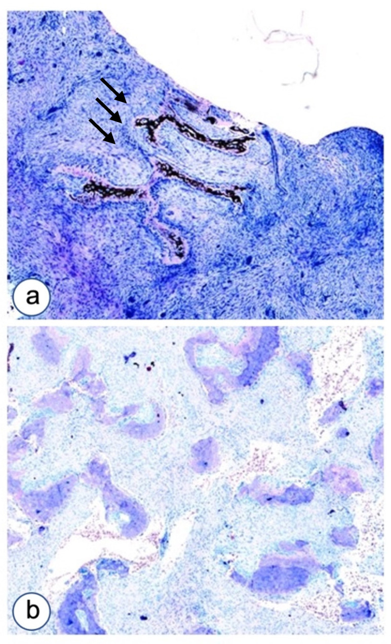
Microscopic aspects of impaired differentiation in Fibrous dysplasia of the jaw as evaluated by Von Kossa–Giemsa histochemical staining. (a) A case with Chinese ideogram aspect and high cellularity composed by fibroblastoid cells committed to osteoblast differentiation associated with giant cells. (b) Another case of FD with curved thin trabeculae of osteoid with high cellularity. Magnifications, ×4 (a,b).