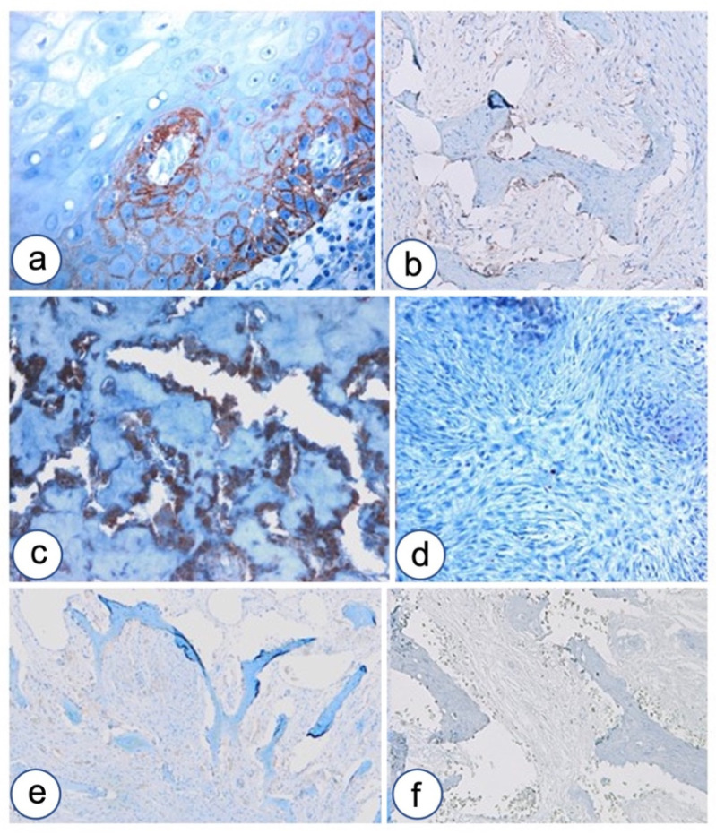
Figure 6.
P-cadherin and E-cadherin expression in FD of the jaw. (a) P-cadherin expression in oral epithelium. (b) P-cadherin expression in remodeling bone surrounding developmental odontogenic cyst. (c) P-cadherin over-expression in pagetoid FD is confined to osteoblasts. (d) P-cadherin negative staining in fibrous area of FD without osteoid formation. (e) E-cadherin shows faint staining in remodeling normal bone. (f) E-cadherin negative staining in FD of the jaw. Magnifications, ×4 (e,f), ×10 (b,c), ×20 (a,d).

