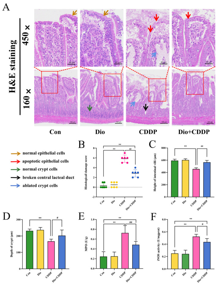
Figure 2.
Ameliorative effect of Dio on ileum tissue injury and inflammatory cell infiltration in rats with CDDP-induced mucositis. (A) Intestinal tissues were stained with H and E dye kits (160×, 450×). Note: Normal epithelial cells (Yellow arrows), apoptotic epithelial cells (Red arrows), normal crypt cells (blue arrows), and Black and blue arrows indicate ablated crypt cells and broken central lacteal duct in cisplatin rats, respectively. (B) Histological damage score. (C) villus length, (D) crypt depth. In addition, determination of MPO (E) and iNOS (F) activity to determine inflammatory infiltration. All values were expressed as Mean ± S.D. (n = 6 samples per group). ** p < 0.01 vs. control group, # p < 0.05, ## p < 0.01 vs. CDDP group.

