Figure 2.
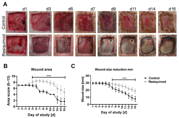
Wound morphology and wound area over the course of the experiment. Wounds of 3 × 3 cm were inflicted on the backs of pigs. Resiquimod was applied for 6 days, and the wounds were further examined over the course of 10 days. (A) Daily visual documentation and scoring was done. The control wounds ((A)–top row) showed physiological wound healing as well as wound contracture. The resiquimod-induced wounds ((A)–bottom row) showed a delay in wound healing and the formation of necrotic scabs. (B) The area scores showed higher values in resiquimod-induced wounds. (C) The wound size was determined by measuring the width of the wound, and less wound size reduction was shown in resiquimod-induced wounds. Data are presented as mean (dot) ± SD (whiskers) (n = 20 for control wounds; n = 40 for resiquimod-induced wounds). Data shown as mean ± SD. Statistical significance has been determined by 2-way ANOVA and corrected for multiple comparisons using the Sidak method. *** (p < 0.001).

