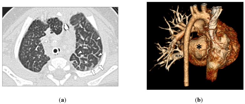
Figure 3.
(a) 3-month-old girl with left-sided pulmonary vein stenosis but no history of aspiration. Axial lung window CT image shows left-sided septal thickening (arrowheads) and pleural thickening (arrows) in the left hemithorax. (b) 3-month-old girl with absent left pulmonary veins due to complete obstruction from severe pulmonary vein stenosis but no history of aspiration. The posterior view of the three-dimensional volume-rendered CT image of the vascular and heart strictures shows absent left pulmonary veins (asterisk). Normal and patent right upper pulmonary vein (RUPV) and right lower pulmonary vein (RLPV) are seen.

