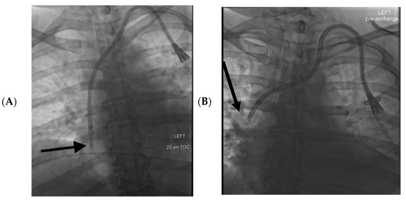
Figure 2.
57-year-old female had a history of end stage renal disease with a left-sided tunneled dialysis catheter. (A): The initial supine fluoroscopic images at the time of catheter placement demonstrated the catheter tip within the mid right atrium (black arrow). (B): Two months later the patient presented with catheter dysfunction, and supine fluoroscopic interrogation demonstrated that the tip of the catheter had migrated 6 cm cranially to the upper SVC (black arrow).

