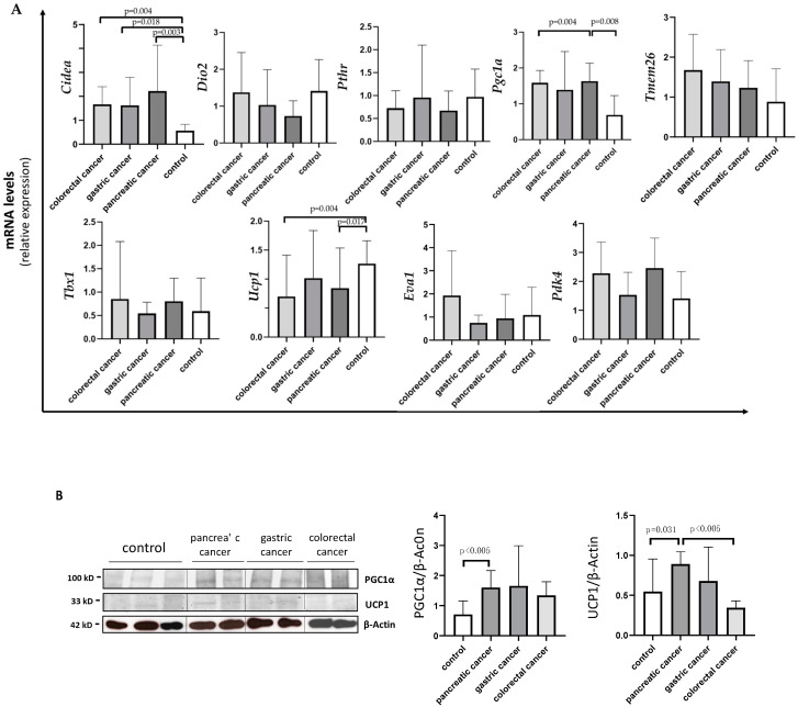
Figure 3.
Evaluation of browning genes and protein expression in white adipose tissue (WAT) of cancer patients divided by the three types of gastrointestinal cancer (colorectal, stomach and pancreas). (A) The mRNA levels of Cidea, Dio2, Pthr, Pgc1α, Tmem26, Tbx1, Ucp1, Eva1 and Pdk4 were analyzed using quantitative real-time PCR in SAT of gastrointestinal cancer patients according to the different type of tumor (gastric N = 7, colorectal N = 8, pancreas N = 9) and in control group (N = 13). (B) Representative Western blot images and protein densitometry quantification for PGC1α and UCP1 in a subset of pancreatic (N = 7), gastric (N = 5) and colorectal (N = 7) cancer patients, and in control group (N = 8). β-Actin was used as loading control.

