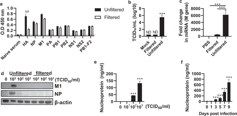
Fig. 1. Release of influenza virus proteins from influenza virus-infected cells.
MDCK cells were infected with the PR8 virus (103 TCID50/ml) for 3 days, followed by filtration using a 50-nm syringe filter. a Internal viral proteins in the culture supernatant were detected by enzyme-linked immunosorbent assay (ELISA). b The viral titer was measured as TCID50/ml. c The M gene was detected by qRT–PCR, and d the M1 and NP proteins were detected by western blotting. e The supernatants of MDCK cells treated with phosphate-buffered saline (PBS) or the PR8 virus (101–103 TCID50/ml) were obtained. After filtration through a 50-nm filter, the nucleoprotein (NP) levels were measured by ELISA. f Mice (n = 4) were infected with the PR8 virus (32 PFU), and then on Days 0, 1, 3, 5, 7, and 9, NP in the bronchoalveolar lavage fluid (BALF) was detected by ELISA. The data in a–c are presented as the mean ± standard deviation (SD) from triplicate culture wells. The data in d are presented as the mean ± standard error of the mean (SEM). ***p < 0.001

