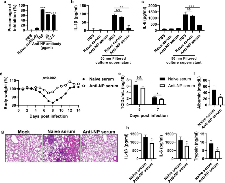
Fig. 6. Anti-NP antibody inhibits the NP-TLR4 interaction and reduces influenza virus pathogenicity.
a An anti-NP antibody was incubated with rNP for 1 h at 25 °C, and then the antibody-treated NP was added to an ELISA plate coated with rTLR4 (2 μg/ml). After sequential incubation and washing, NP bound to rTLR4 was detected by ELISA. b, c MDCK cells were infected with the PR8 virus (103 TCID50/ml) for 72 h, and the cell culture supernatant was filtered using a 50-nm syringe filter. The filtered supernatant was incubated with naïve serum (1:100 diluted) or anti-NP serum (1:100 diluted). Subsequently, BMDMs were treated with the filtered supernatant for 20 h, and the levels of cytokines and chemokines in the cell culture supernatants were measured by ELISA. Mice (n = 6) were infected with the PR8 virus (32 PFU) and then injected with anti-NP serum (200 μl/mouse) intraperitoneally daily for three days. d Body weight was monitored for two weeks. e The TCID50 was evaluated using BALF samples obtained on Days 5 and 7 post-infection. f, g The albumin level in the BALF was measured, and the histology of lung tissue was assessed by H&E staining at 5 days post-infection. h Proinflammatory cytokines and trypsin in the BALF were measured by ELISA at five days post-infection. The data in a are presented as the mean ± SD from triplicate culture wells. The data in b–f are presented as the mean ± SEM. ***p < 0.001, **p < 0.01, *p < 0.05

