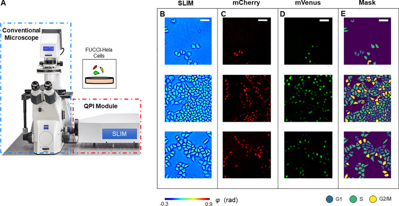
Figure 1.
Schematic of the imaging system. (A) The SLIM module was connected to the side port of an existing phase contrast microscope. This setup allows us to take colocalized SLIM images and fluorescence images by switching between transmission and reflection illumination. (B) Measurements of HeLa cells. (C) mCherry fluorescence signals. (D) mVenus fluorescence signals. (E) Cell cycle stage masks generated by using adaptive thresholding to combine information from all three channels. Scale bar is 100 μm.

