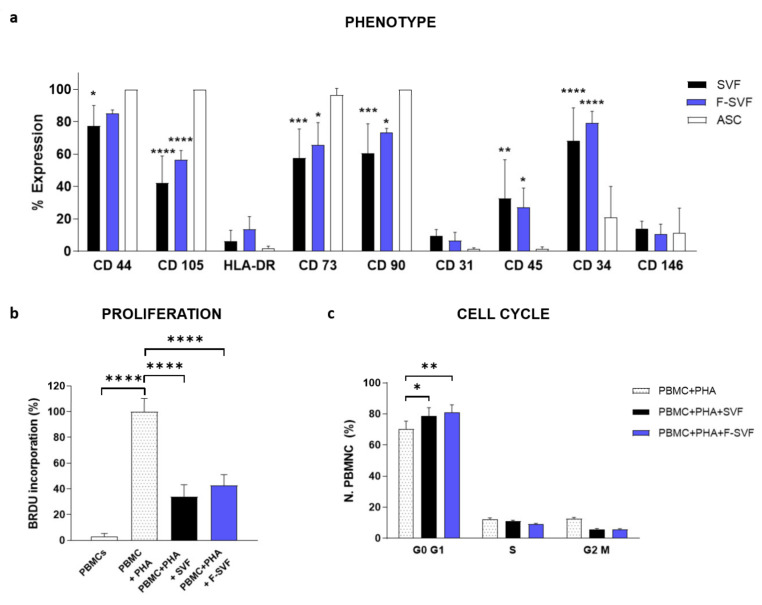
Figure 5.
Phenotype characterization of stromal vascular fraction (SVF) and the fraction derived stromal vascular fraction (F-SVF) cells by flow cytometry. Mesenchymal markers were slightly more expressed in the F-SVF, proving an enrichment of the mesenchymal component of the SVF due to the label-free sorting (a). SVF and F-SVF cells showed the same immunomodulatory ability to inhibit the proliferation of PHA-stimulated PBMCs by BrdU assay (b). Moreover, it was shown that SVF and F-SVF cells arrested stimulated PBMCs in the G0/G1 phase of the cell cycle (c). *: p < 0.05; **: p < 0.01; ***: p < 0.001; ****: p < 0.0001.

