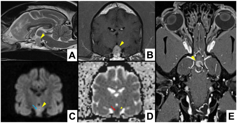
Figure 3.
Dog 1: A total of 3 Tesla MRI scan of a pituitary mass (yellow arrowheads), showing hyperintense signal in DWI (light blue arrow) and hypointense in the corresponding ADC map (red arrow), suggesting acute pituitary haemorrhage, heterogeneous contrast enhancement with an hypointense area (black arrow). (A) T2W in sagittal plane as reference position; (B) T1W post-contrast; (C) DWI b1000; (D) ADC map transverse sections; (E) T1W dorsal section.

