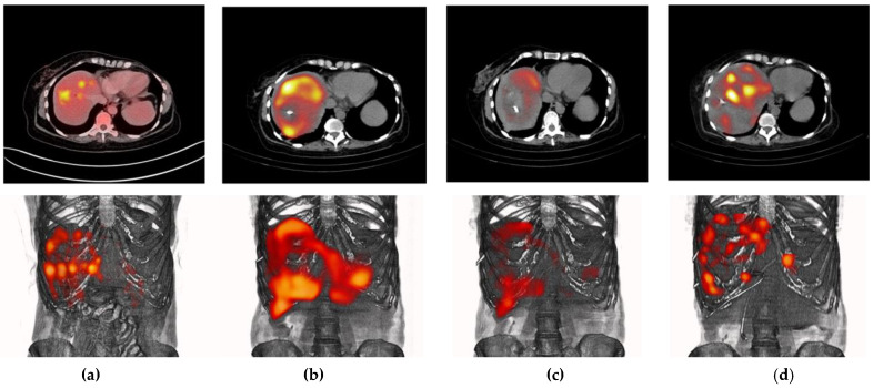
Figure 5.
Different molecular images (top: axial sections; bottom: PET/CT or SPECT/CT images) of a patient with multiple colorectal liver metastases (PSMA-positive): (a) [18F]FDG PET/CT images acquired on 27 August 2021; [99mTc]Tc-iPSMA SPECT/CT images at (b) 3 h and (c) 24 h post-injection (administration on 9 November 2021); and (d) 177Lu2O3-iPSMA SPECT/CT images acquired on 18 November 2021. It can also be observed that over almost three months, the metastases moved due to the appearance of bilomas and the presence of necrotic tumors. The 177Lu2O3-iPSMA SPECT/CT image was contrast-enhanced to highlight the uptake of the radionanoparticles in tumor lesions as proof of concept. The detailed biokinetic behavior of the source organs is shown in Figure 6.

