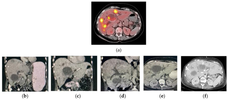
Figure A1.
(a) PET/CT liver axial image of a patient with liver metastases of colorectal cancer (August, 2021) showing a necrotic mass without metabolic activity (approximate size of 6 cm on its major axis). (b–f) Different CT images of the patient acquired in November 2021. Observe the hepatomegaly progression, the presence of different bilomas, and dilatation of the intrahepatic bile ducts.

