Figure 2.
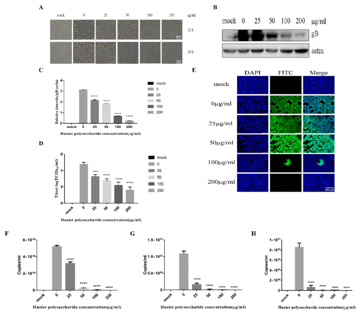
Huaier polysaccharide inhibits PRV XJ5 infection in PK15 cells. (A) Infected cells treated with 25, 50, 100, or 200 μg/mL Huaier polysaccharide for 12 or 24 h, showing changes in cell morphology. (B) gB and actin protein expression was determined through Western blot assay. (C) The relative intensity of intracellular gB to that of actin. Data are presented as means from three independent statistical experiments. The relative intensities of protein were quantified using Image J. Significance was analyzed using a one-tailed Student’s t-test. (D) The viral titers were evaluated through 50% tissue culture infective dose (TCID50), and (E) immunofluorescent assay (IFA) for internalized virus was performed. (F–H) PRV gB were assessed with qRT-PCR analysis in PK15 cells treated with Huaier polysaccharide (25, 50, 100, and 200 μg/mL) at 24 h.p.i. *** p = 0.0003, **** p < 0.0001. Data are shown as mean ± SD based on three independent experiments.

