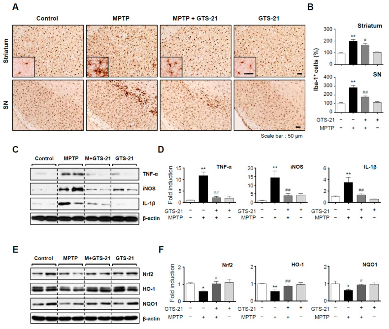
Figure 8.
Effect of GTS-21 on microglial activation, inflammatory markers, and antioxidant enzyme expression in the brains of MPTP-injected mice. (A) Iba-1-positive microglial cells in the substantia nigra and striatum (representative images). (B) Quantitative analysis was performed by measuring the number of Iba-1-positive cells (n = 4 per group, 3 sections/brain). (C–F) Protein extracts from the substantia nigra of each group were analyzed using TNF-α, iNOS, and IL-1β antibodies or Nrf2, HO-1, and NQO1 antibodies (n = 4). The figures show representative blots (C,E) and quantification data (D,F). The data are presented as the mean ± SEM. * p < 0.05, vs. control group; ** p < 0.01, vs. control group; # p < 0.05 vs. MPTP-treated group; ## p < 0.01 vs. MPTP-treated group.

