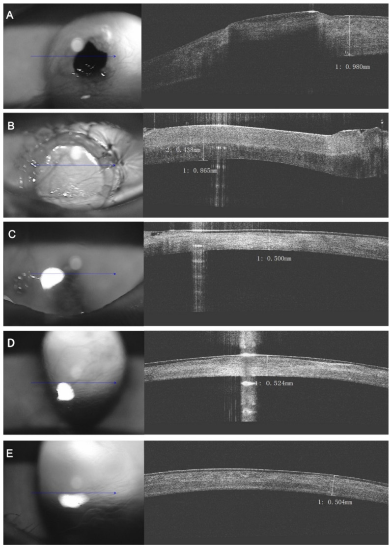
Figure 6.
The same patient as Figure 1. Optical coherence tomography (OCT) upon the eye. (A) Preoperative image; (B) Immediate examination after acellular bioengineering cornea lamellar keratoplasty: The fine interface between the graft and the recipient side; (C) Three months post-operatively: No obvious transition between the graft and the recipient side, both of which were stable; (D) Six months post-operatively: The whole cornea was showed highly integral and uniform; (E) Twelve months post-operatively: The alignment of the collagen fibers in the whole cornea was integrated and restored. (Blue arrows: corneal L-Scan pattern).

