Figure 1.
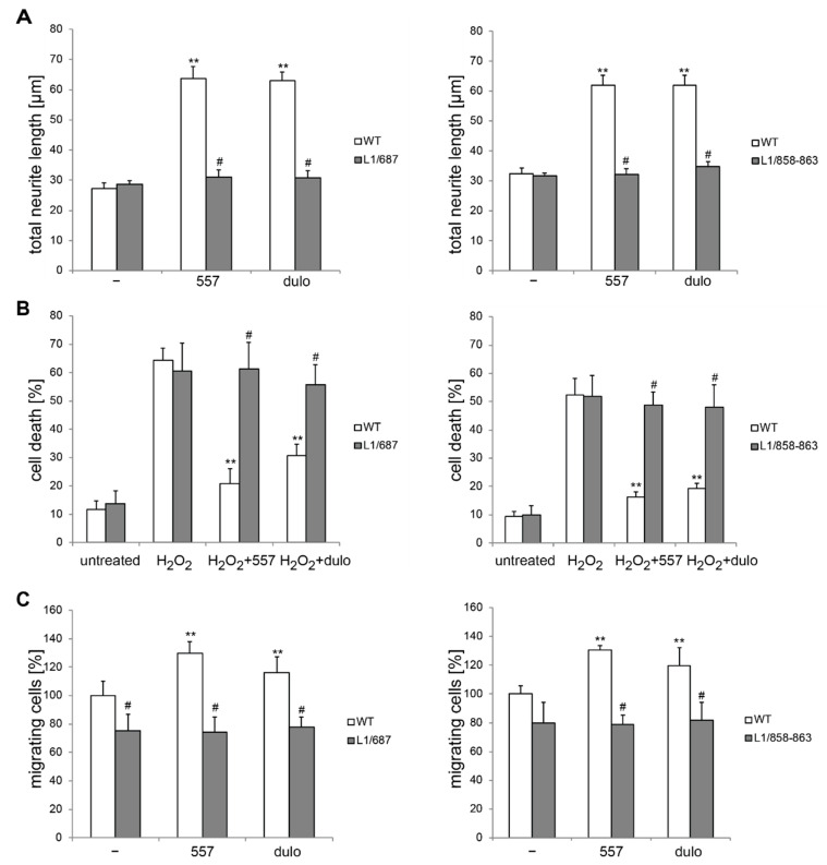
Cerebellar neurons from L1/858–868 and L1/687 mice do not respond to treatment with antibody 557 or duloxetine. Cerebellar neurons and explants were prepared from 6- to 7-day-old male wild-type (WT, white bars) and L1 mutant littermate (gray bars) mice and maintained on PLL substrate. (A) Neurons were left untreated (−) and treated with 50 µg/mL of antibody 557 (557) or 100 nM of duloxetine (dulo) for 24 h, and neurite outgrowth was determined from 100 cells per genotype and condition for each experiment. (B) Cell death was measured in the absence (untreated) or presence of 10 µM hydrogen peroxide (H2O2) and 50 µg/mL of antibody 557 (H2O2+557) or 100 nM of duloxetine (H2O2+dulo) for 24 h. Cells were stained with calcein and propidium iodide, and cell death was measured by counting live and dead cells from three wells per genotype and treatment. (C) After 16 h of culturing, explants were left untreated (−) or treated for 24 h with 50 µg/mL of antibody 557 (557) or 100 nM of duloxetine (dulo). Migrating cells from 12 explants per condition and genotype were counted. (A–C) Values show the means ± SD from three (B,C) or four (A) independent experiments. ** p < 0.01 difference relative to PLL, # p < 0.05 difference relative to the wild-type (two-way ANOVA with Tukey’s post-hoc test).

