Figure 6.
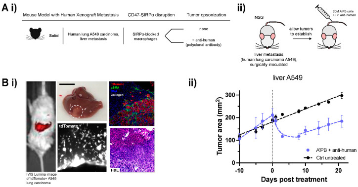
A’PB therapy represses growth in human metastatic WT CD47 tumors. (A) (i) Table of human xenograft mouse model details. Human lung A549 carcinoma is surgically implanted at an orthotopic metastatic site in the livers of NSG mice. Anti-SIRPα is used to block CD47 signaling on adoptively transferred marrow cells and anti-human is the source of tumor-opsonizing antibody (A’PB). (ii) Schematic of tumor induction and treatment. NSG mice were surgically implanted with liver metastases. Mice were treated with A’PB marrow on the indicated day 0, and 3 times weekly for 2 weeks with anti-human i.v. (B) (i) Representative whole-body fluorescence image of tdTomato signal in surgically implanted liver metastases (left). Images of explanted liver with liver metastasis (middle top, A549 tumor circled, scale: 1 cm) and tdTomato signal of A549s infiltrating stroma (middle bottom). (ii) Sectioned liver tissue images showing tdTomato, αSMA, DNA, and collagen of A549 metastases relative to normal liver tissue (top right) and H&E staining (bottom right). (ii) In vivo growth curve of liver metastatic human lung adenocarcinoma A549 tumors. A’PB and tumor-opsonizing antibody repress tumor growth by 50% over several days relative to untreated control. Tumor burden is monitored by fluorescent imaging with an IVIS Lumina II (fluorophore: tdTomato). Without additional treatment to continue tumor cell clearance, metastases begin to expand again after about 10 days. Error bars represent s.e.m. Exponential decay for Tumor area in the regression phase of the treated tumor fits to exp(−t/1.55) for 0 < t < 8.

