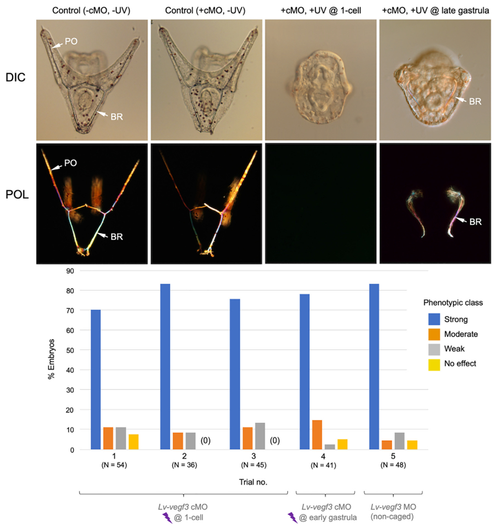
Fig. 4.

In vivo analysis of Lv-vegf3 cMO. The top two rows of panels show living embryos (24 hpf) viewed with differential interference contrast (DIC) and polarization (POL) optics. Control embryos (−cMO, −UV and +cMO, −UV) developed extensive, branched skeletons that contained elongated, paired body rods (BR) and postoral rods (PO). Decaging of the Lv-vegf3 cMO at the 1-cell stage (+cMO, +UV @ 1-cell; aboral view) completely blocked skeleton formation in most embryos, phenocopying the effects of the non-caged form of the MO. Decaging at the late gastrula stage (16 hpf) (+cMO, +UV @ late gastrula; blastoporal view) blocked the growth of postoral rods but not that of body rods, mimicking the effect of treating late gastrula stage embryos with axitinib, a highly specific inhibitor of Vegf signaling (Adomako-Ankomah and Ettensohn, 2013). Bottom panel- Quantification of phenotypes of embryos injected with Lv-vegf3 cMO and irradiated at the 1-cell (Trials 1–3) or early gastrula (12 hpf; Trial 4) stages, or injected at the 1-cell stage with the non-caged form of the same MO sequence (Trial 5). Morphant phenotypes were scored as shown in Fig. 3.
