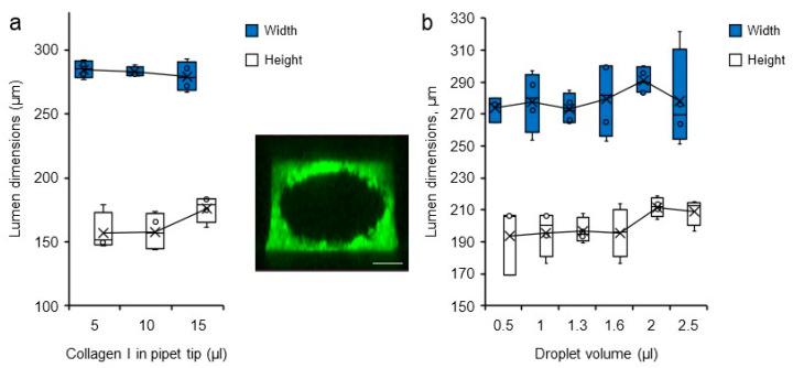
Figure 4.
Variations in lumen dimensions. Dimensions (height and width) of the lumens measured by confocal microscopy according to (a) the volume of collagen I solution loaded in the pipet tip at the time of channel filling n = 4, and (b) the volume of droplets loaded into the microchannel filled with collagen solution, n = 4. (insert). Confocal microscopy maximum intensity projection 3D reconstruction of a representative collagen I/BSA-FITC channel used for measurements, bar = 200 µm.

