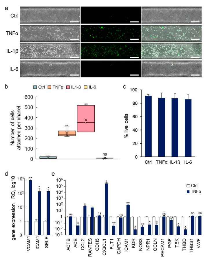
Figure 9.
Immune activation of VoC. (a) VoC were stimulated or not with TNFα (0.57 nM), IL-1β (0.59 nM), or IL-6 (0.48 nM) for 4 h, then, calcein AM-labeled THP-1 monocytes (green) were injected in the lumens and allowed to adhere onto the luminal face of the endothelium. Left—phase contrast; middle—fluorescent imaging; right—merged images, bar= 200 µm. (b) Quantification of the numbers of immune cells attached to the vessel walls along the entire channels, n = 3. (c) Percentage of living endothelial cells after treatments with cytokines as in (a). (d) RT-qPCR array analysis of gene expression in endothelial cells recovered from VoC following stimulation or not with TNFα (0.57 nM) for 4 h. (e) Taqman array card analysis of expression of genes following extraction of single vessels-on-chips, a selection of genes is presented, n = 4, * p < 0.05, ** p < 0.01; ns—nonsignificant, compared to Ctrl.

