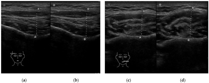
Figure 1.
Example of an ultrasound examination of muscle thickness. (a) The temporalis muscle in the relaxed mandible position with slight contact between opposing teeth; (b) the temporalis muscle in the maximum voluntary clenching; (c) the masseter muscle in the relaxed mandible position with slight contact between opposing teeth; (d) the masseter muscle in the maximum voluntary clenching; line A-A—measuring line of the thickness of the muscle in the relaxed mandible position with slight contact between opposing teeth; line B-B—measuring line of the thickness of the maximum voluntary clenching.

