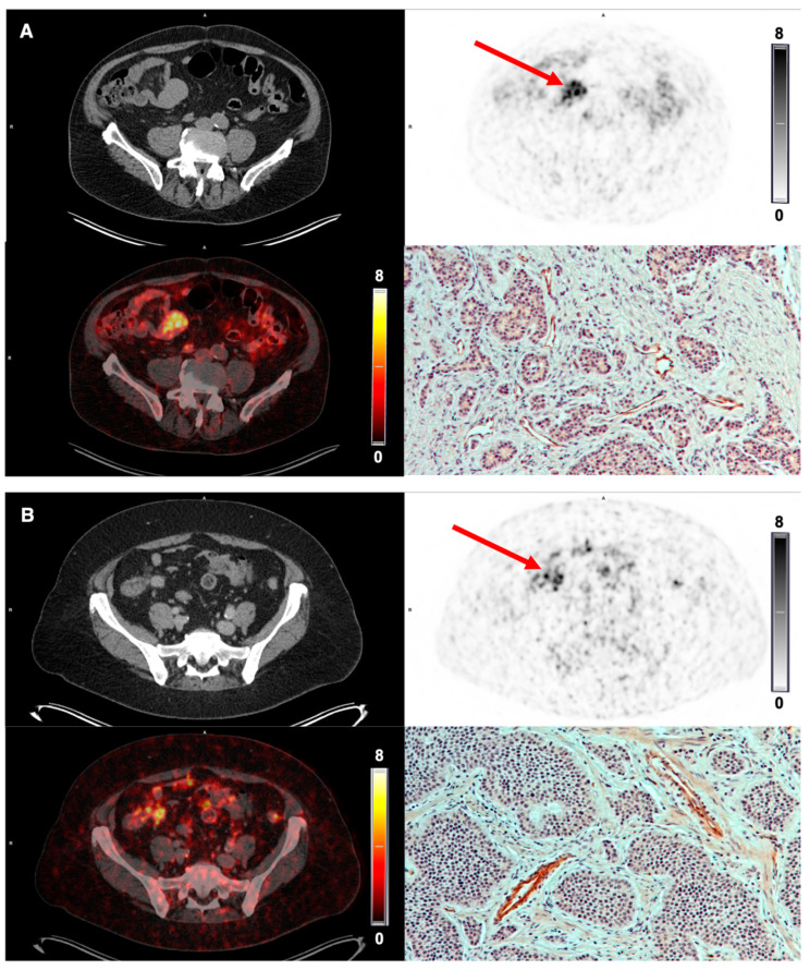
Figure 6.
[68Ga]Ga-NODAGA-E[c(RGDyK)]2 PET imaging in NET. (A) Representative transverse CT, PET (1 h p.i.) and fused PET/CT images of primary tumor lesion (red arrow) with a high uptake of [68Ga]Ga-NODAGA-E[c(RGDyK)]2 in small intestine primary tumor (patient 2) and high intensity immunohistochemistry staining for integrin αvβ3. (B) Patient 3 also displays a high uptake of tracer in the terminal ileum primary tumor and a corresponding high intensity of integrin αvβ3 immunohistochemistry staining.

