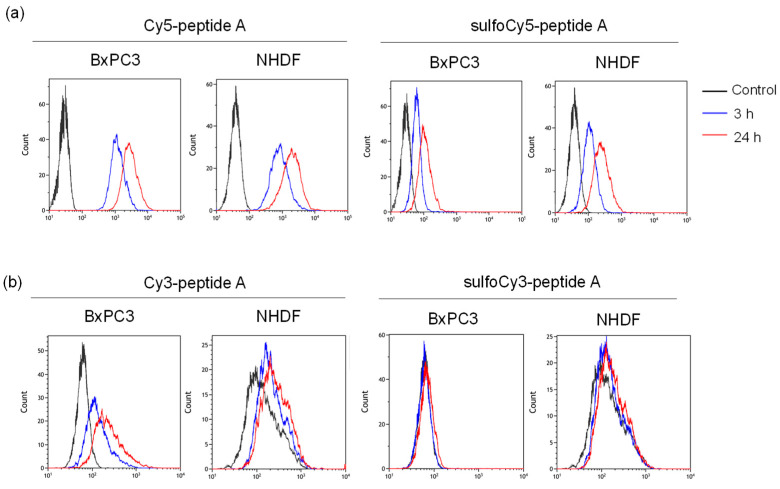
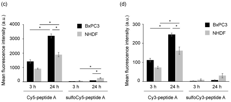
Figure 4.
Flow cytometry of BxPC3 and NHDF cells treated with dye-peptide A. The histograms of (a) Cy5/sulfoCy5-peptide A and (b) Cy3/sulfoCy3-peptide A. The analysis of (c) Cy5/sulfoCy5-peptide A and (d) Cy3/sulfoCy3-peptide A. Cells were incubated with 0.1 μM dye-peptide A at 37 °C for 3 or 24 h before flow cytometry. The data represent the mean ± SEM (n = 3). The statistical significance was assessed by Tukey-Kramer test (* p < 0.01).


