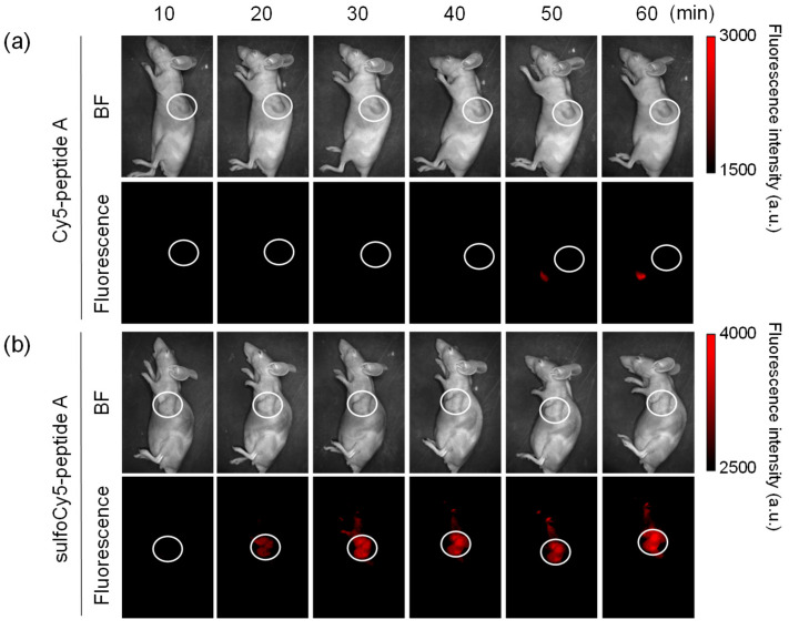
Figure 5.
In vivo fluorescence imaging of tumor-bearing mice injected with dye-peptide A. The tumor-bearing mice were intravenously given 0.1 mL of (a) 100 μM Cy5-peptide A and (b) 100 μM sulfoCy5-peptide A. Ten minutes after the injection, the images were taken from a side view. The tumor is indicated by white circles. The in vivo experiment was performed three times.

