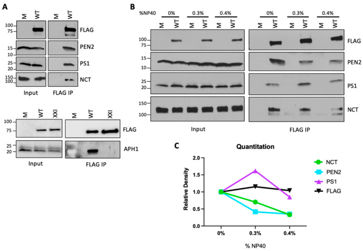
Figure 1.
Characterizing the interaction between HPV and γ-secretase. (A) HeLa cells were treated with 1 µM XX1 γ-secretase inhibitor as indicated (bottom panels) or left untreated, then infected at MOI 50 with HPV16 PsV containing a 3XFLAG tag on the C-terminus of L2 (WT) or mock (M) infected. Cell lysates were collected 16 h.p.i. in lysis buffer containing 1% DMNG. L2 was immunoprecipitated with anti-FLAG, and samples were analyzed by Western blotting for HPV L2 (FLAG) and the indicated γ-secretase subunit. The inputs were total cell lysates without immunoprecipitation. Similar results were obtained in eight independent experiments. (B) HeLa cells were infected, immunoprecipitated, and analyzed as in (A), except the lysis buffer containing the indicated percentage of NP40 was supplemented with DDM to make the total detergent concentration 1%. Similar results were obtained in three independent experiments. (C) The ImageJ Gel Analysis tool was used to quantitate the relative density of each band in (B), normalized to the density of the respective band in samples lysed in 1% DDM (set to a relative density of 1).

