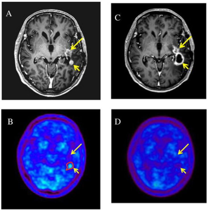
Figure 3.
Two enhanced lesions (long and short arrow) were demonstrated in the left temporal lobe on T1-weighted magnetic resonance imaging (A), MET-PET demonstrated a MET high-uptake on the region of short arrow (B), only the enhanced lesion (short arrow) was treated with RT; 5 months later it was increased in size (C) but not in uptake (D) (suggestive of pseudoprogression) while the non treated lesion remained stable [58].

