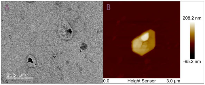
Figure 3.
TEM and AFM imaging of exosomes isolated by aptamer-based microfluidics. (A) TEM image of EVs isolated by microfluidics presents a distorted cup-shaped morphology and uniform unimodal size distribution following 200 nm filtration. (B) AFM image EVs isolated by microfluidics presents a distorted cup-shaped morphology.

