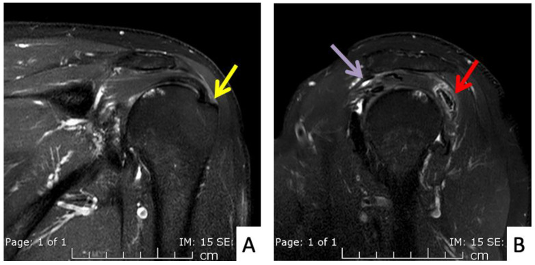
Figure 5.
MRI images of the left shoulder of Patient 4; (A) coronal and (B) sagittal T2-weighted images of the left shoulder reveal a small, partial-thickness, bursal surface tear at the footprint of the supraspinatous tendon (yellow arrow). Increased signal intensity of the myotendinous junction of the infraspinatous tendon indicates muscle strain (red arrow). An edematous subacromial bursa with thin fluid (purple arrow) was also observed.

