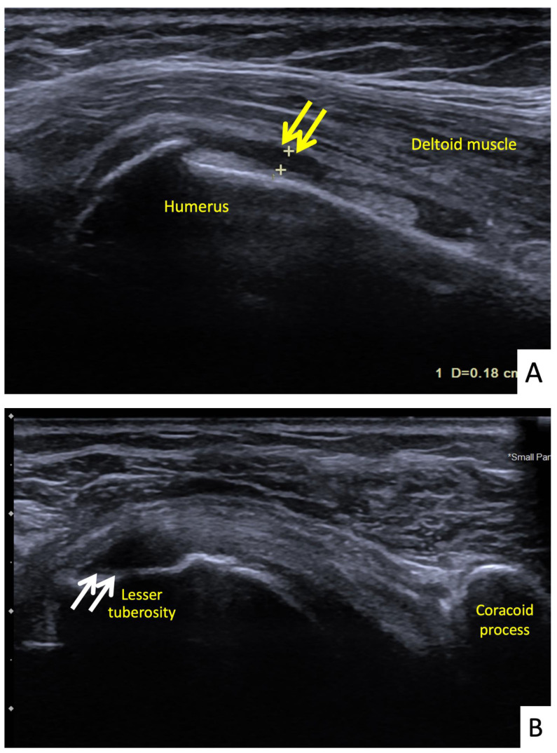
Figure 6.
Ultrasonographic images of the right shoulder of Patient 5. (A) A longitudinal ultrasonographic image over the lateral aspect of the left proximal humerus with the patient in supination showing a small amount of fluid (yellow arrows) in the mildly distended subdeltoid bursa. (B) A transverse ultrasonographic image over the lesser tuberosity of the right shoulder with the patient in the external rotation position showing a partial thickness, articular surface tear at the footprint of the subscapularis tendon (white arrows).

