Figure 2.
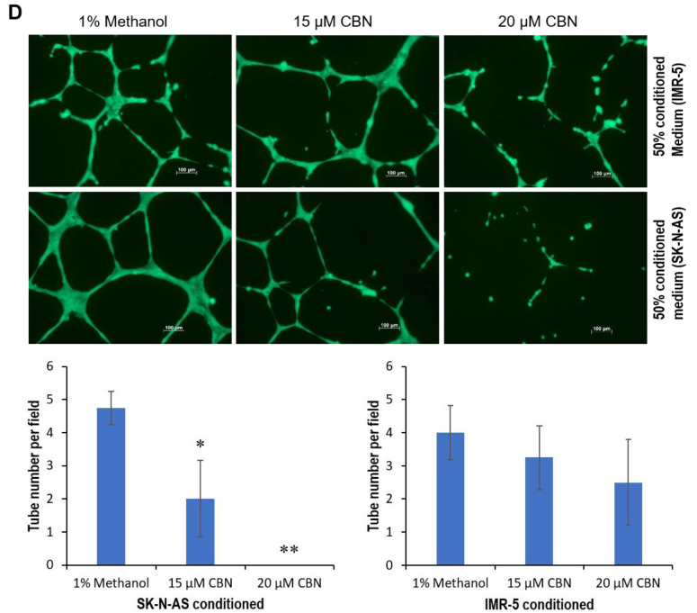
Anti-neuroblastoma effect of CBN via inhibition of AKT pathway and transactivation of miR-34a. (A) 1000 IMR-5 cells per well and 2000 SK-N-AS cells per well were plated in 96-well plates. At 24 h after plating, cells were treated with the indicated concentration of CBN, and MTT assay was performed using Cell Proliferation Kit I as described in Section 4. (B,C) IMR-5 and SK-N-AS cells grown to 85% confluency were exposed to either 15 or 20 μM CBN. At 48 h after treatment, cells were harvested for cell cycle and apoptosis analyses. (D) Tube formation assay was carried with 50% of conditioned medium from either IMR-5 or SK-N-AS cells exposed to either 15 or 20 µM CBN as detailed in Section 4; representative images were taken using a fluorescence microscope (100×). Data were expressed as mean ± SD for five images. (E) Cell invasion assay was performed using 33% of conditioned medium from either IMR-5 or SK-N-AS cells exposed to either 15 or 20 µM CBN as detailed in Section 4; representative images were taken under inverted microscope (200×). Data were expressed as mean ± SD for five images. (F) IMR-5 and SK-N-AS cells grown to 85% confluency were exposed to either 15 or 20 μM CBN. At 48 h after treatment, total RNA was isolated and subjected to qRT-PCR analysis using hsa-miR-34a primer set. (G,H) IMR-5 and SK-N-AS cells grown to 85% confluency were exposed to either 15 or 20 μM CBN. At 48 h after treatment, whole cellular lysates were prepared and subjected to Western blotting with antibodies to CBR2, pAKT1/2/3, AKT1, pERK1/2, ERK1/2, CDK2, cyclin E1, E2F1, notch1, snail, and p53, GAPDH served as a loading control. (I) IMR-5 cells grown to 80% confluency were transfected with either 10 nM LNA miR-34a Power Inhibitor or 10 nM negative control A. At 24 h after transfection, 2000 cells per well were plated in 96-well plates and exposed to 15 μM CBN; MTT assay was carried out as described in Section 4. (J) IMR-5 cells grown to 80% confluency were transfected with either 10 nM LNA miR-34a Power Inhibitor or 10 nM negative control A. At 24 h after transfection, cells were exposed to 15 µM CBN. At 48 h after exposure, whole cellular lysates were prepared and subjected to Western blotting with antibody against E2F1; GAPDH served as a loading control. (K) 3000 BJ-5ta cells per well and 5000 WI-38 cells per well were plated in 96-well plates. At 24 h after plating, cells were exposed to the indicated concentration of CBN, and the MTT assay was performed using Cell Proliferation Kit I as detailed in Section 4. *, p < 0.05; ** p < 0.01, ***, p < 0.001. Original western blot data is shown in Figure S3.


