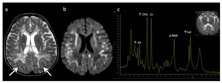
Figure 7.
Metachromatic leukodystrophy in a 16-month-old female who presented with loss of developmental milestones. (a) Axial T2WI and (b) axial DWI demonstrate cerebral volume loss with ex vacuo ventriculomegaly, a widespread leukodystrophy with increased white matter signal sparing the U-fibers (arrows) and involving corpus callosum, and reduced diffusion at leading-edges of active demyelination. (c) SV-MRS over the left periatrial white matter shows elevated Lac, severely depleted NAA, and slightly elevated Cho and mI. Findings are consistent with considerable axonal damage and loss of the white matter.

