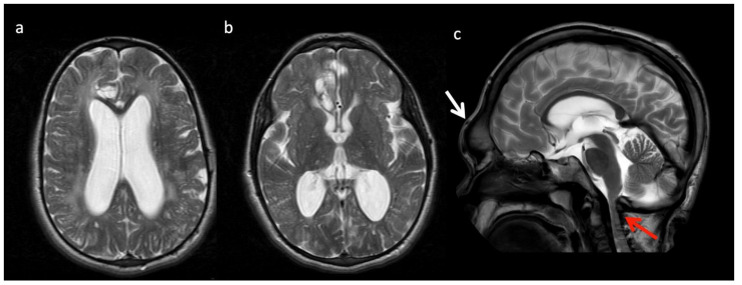
Figure 11.
Thirteen-year-old male with Hurler’s disease (mucopolysaccharidosis). (a,b) Axial T2W at the level of the corona radiata (a) and third ventricle (b) show diffuse white matter hyperintensity, multiple enlarged perivascular spaces, and generalized ventriculomegaly. Volume loss in the right frontal lobe may be due to prior injury. (c) Sagittal T2 images from a different 13-year-old male with Hurler’s shows frontal bossing (white arrow), dens hypoplasia, platyspondyly, J-shaped sella, and thickened dural ring at the foramen magnum with craniocervical junction (CVJ) stenosis (red arrow).

