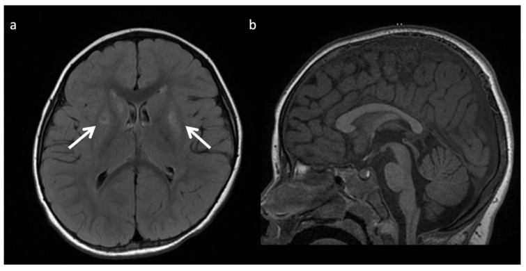
Figure 16.
Pyruvate dehydrogenase complex (PDHc) deficiency in a 30-month-old female with desaturations. (a) Axial T2 FLAIR image shows a Leigh pattern of brain injury with bilateral putamen and right caudate head (white arrows) hyperintense lesions with central signal suppression representing necrosis. (b) Sagittal T1WI shows mild pontine hypoplasia and thinning of the callosal splenium, likely representing regional hypogenesis rather than volume loss, given low normal callosal length and normal cerebral white matter depth.

