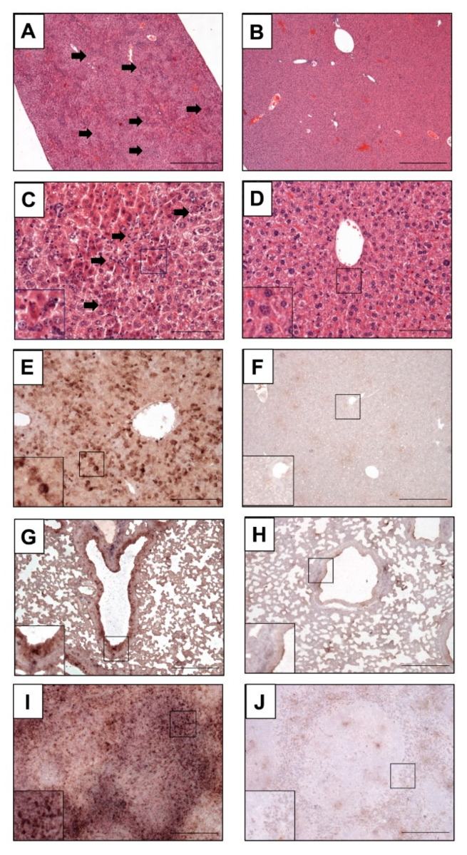
Figure 5.
Histopathological examination of infected mice. (A,C,E,G,I) Mock-vaccinated control mice (PBS) compared to MVA-EBOV-NP-vaccinated mice (B,D,F,H,J); (A–D) H&E staining, (G–J) in situ hybridization with digoxigenin-labeled RNA probes binding to EBOV GP mRNA, brown staining; (A–F): liver, (G,H): lung, (I,J): spleen. Livers of PBS mock-vaccinated mice (A,C) showing randomly distributed hepatocellular necrosis and lymphohistiocytic infiltrations (arrows). Insert: magnification of selected areas; Scale bar (A,B): 500 µm; (C,D): 100 µm; (E–J): 200 µm.

