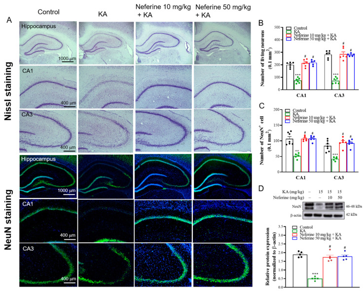
Figure 2.
Results of Nissl and NeuN staining in the rat hippocampus. (A) Representative images and (B,C) quantitative data for the number of Nissl- or NeuN-positive hippocampal neurons (n = 5–7 rats/group). Neferine increased the numbers of Nissl- or NeuN-positive hippocampal neurons in the CA1 and CA3 areas (one-way ANOVA). (D) Representative Western blot images in the different groups and densitometric values for NeuN were normalized to β-actin levels (n = 5 rats/group). Statistical results of immunoblot analysis show that neferine increased the band intensities of NeuN (one-way ANOVA). ** p < 0.01. *** p < 0.05 vs. control group. # p < 0.05 vs. KA-treated group.

